Quantitative ATP Analysis
Quantitative ATP Analysis
Achieving precision in quantitative ATP analysis is essential for accurately assessing cellular energy status and metabolic activity. Tribioscience’s ATP Colorimetric and Fluorometric Assay Kit (Catalog #TBS2010) offers a robust and straightforward method for detecting ATP concentrations as low as 0.5 µM in various biological samples. The assay employs the phosphorylation of glycerol to generate a product that can be quantified either colorimetrically (OD = 570 nm) or fluorometrically (Ex/Em = 535/590 nm), providing flexibility based on the sensitivity requirements of the experiment. The kit’s design ensures high sensitivity and accuracy, utilizing only 10 µL of sample per test and covering a detection range of 0.5–1000 µM in a 96-well plate format. The entire procedure is streamlined, taking less than 30 minutes to complete, which facilitates high-throughput analysis without compromising precision.
The assay protocol emphasizes the importance of sample preparation to maintain ATP integrity. For accurate measurements, tissue samples (1–10 mg) or cells (1 × 10⁶) should be lysed in 100 µL of assay buffer. To preserve ATP levels, samples intended for later analysis should be rapidly frozen using liquid nitrogen or dry ice. After lysis, samples are centrifuged at 15,000 × g for 2 minutes at ice-cold temperatures to remove insoluble materials, and the supernatant is used for the assay. The reaction mix, comprising assay buffer, substrate, probe, and enzyme, is added to the samples, followed by incubation at room temperature for 20 minutes protected from light. The resulting signal is then measured, and ATP concentrations are calculated by comparing the sample readings to a standard curve generated using known ATP concentrations.
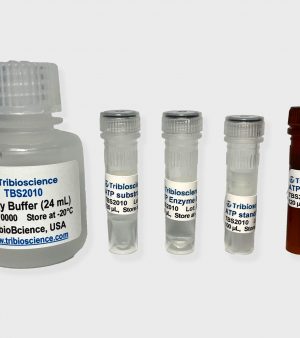
The kit’s components are designed for stability and ease of use. It includes assay buffer, probe, substrate, enzyme, and a 50 mM ATP standard, all of which should be stored at –20°C to maintain their efficacy. Before use, all components except the enzyme should be brought to room temperature, and small vials should be briefly centrifuged prior to opening to ensure complete reagent recovery. The assay’s design allows for both colorimetric and fluorometric detection methods, with the fluorometric approach offering 10–100 times higher sensitivity than the colorimetric method. This versatility makes the kit suitable for a wide range of applications, including direct ATP assays in cells and tissues, as well as the analysis of enzymes that produce or degrade ATP.